FineTest
SKU(재고 관리 코드):FNab10248
anti- MAVS antibody
anti- MAVS antibody
Delve into the intricate world of antiviral defense and innate immunity research with our Anti-MAVS Antibody. Available in a 100µg size, this antibody is your indispensable tool for understanding the role of Mitochondrial Antiviral-Signaling Protein (MAVS) in orchestrating immune responses to viral infections. Key Features: Precise Targeting: This antibody is rigorously validated for specific recognition of MAVS, ensuring accurate and reliable results in your studies of antiviral defense, immune signaling, and host-pathogen interactions. Versatile Applications: Compatible with a variety of research techniques, including Western blotting, immunoprecipitation, and immunofluorescence. It's ideal for researchers studying viral infections, innate immunity, and antiviral therapies. High Sensitivity: The Anti-MAVS Antibody is optimized for superior sensitivity, enabling you to detect low-abundance MAVS in complex cellular samples, facilitating a deeper understanding of antiviral signaling pathways. Broad Research Impact: From unraveling antiviral responses to exploring host defense mechanisms, this antibody is a valuable asset for researchers in virology, immunology, and infectious disease research. Incorporate the Anti-MAVS Antibody into your research to gain deeper insights into the role of MAVS in immune responses to viral infections. Its precision and reliability make it an indispensable asset for scientists and researchers in diverse fields.
Product Name
MAVS antibody
Size
100µg
Form
liquid
Purification
Immunogen affinity purified
Purity
≥95% as determined by SDS-PAGE
Host
Rabbit
Clonality
polyclonal
Isotype
IgG
Storage
PBS with 0.02% sodium azide and 50% glycerol pH 7.3, -20℃ for 12 months(Avoid repeated freeze / thaw cycles.)
BACKGROUND
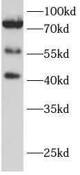
Required for innate immune defense against viruses. Acts downstream of DDX58/RIG-I and IFIH1/MDA5, which detect intracellular dsRNA produced during viral replication, to coordinate pathways leading to the activation of NF-kappa-B, IRF3 and IRF7, and to the subsequent induction of antiviral cytokines such as IFN-beta and RANTES(CCL5). Peroxisomal and mitochondrial MAVS act sequentially to create an antiviral cellular state. Upon viral infection, peroxisomal MAVS induces the rapid interferon-independent expression of defense factors that provide short-term protection, whereas mitochondrial MAVS activates an interferon-dependent signaling pathway with delayed kinetics, which amplifies and stabilizes the antiviral response. May activate the same pathways following detection of extracellular dsRNA by TLR3. May protect cells from apoptosis.It can undergoe phosphorylation on multiple sites and ubiquitination, which may together cause the molecular weight migrate to about 70 kDa despite the predicated 57 kDa.
IMMUNOGEN INFORMATION
Immunogen
mitochondrial antiviral signaling protein
Synonyms
CARD adapter inducing interferon beta,Interferon beta promoter stimulator protein 1,Cardif,Putative NF-kappa-B-activating protein 031N,Virus-induced-signaling adapter
Observed MW
40kDa,56kDa,70kDa
APPLICATION
Tested Application
ELISA, WB, IHC
Recommended Dilution
WB: 1:500-1:2000;IHC: 1:50-1:500
UNIPROT INFORMATION
UniProt ID
IMAGES
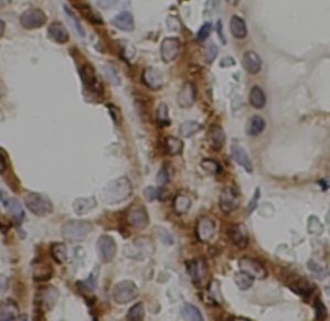
Immunohistochemistry of paraffin-embedded mouse brain tissue slide using FNab10248(MAVS Antibody) at dilution of 1:100
rat kidney were subjected to SDS PAGE followed by western blot with FNab10248(MAVS antibody) at dilution of 1:1000