FineTest
SKU(재고 관리 코드):FNab00129
anti- ACVR1B antibody
anti- ACVR1B antibody
Investigate the role of ACVR1B in signaling pathways with our Anti-ACVR1B Antibody. This indispensable research tool, available in a generous 100µg size, provides you with the precision and reliability needed to explore ACVR1B and its involvement in cellular signaling.
ACVR1B, also known as activin receptor type-1B, is a member of the TGF-β receptor family and is implicated in various signaling cascades. It plays a crucial role in multiple cellular processes, including cell differentiation and tissue development. Our antibody is precision-engineered to specifically target ACVR1B with remarkable precision and sensitivity. Rigorously validated, it ensures consistent and dependable results, instilling confidence in your exploration of signaling pathways.
Produced using state-of-the-art techniques, this antibody is sourced from rabbit hosts, offering a polyclonal clonality that broadens its range of binding capabilities. Its versatility makes it an ideal choice for diverse applications, including Western blotting, immunoprecipitation, and immunofluorescence assays.
Whether you're unraveling the roles of ACVR1B in cell differentiation, studying tissue development, or investigating ACVR1B-related conditions, our Anti-ACVR1B Antibody is your trusted research companion. Its high-quality formulation and generous 100µg size ensure it is an indispensable asset for your laboratory, enabling groundbreaking discoveries and contributions to cellular signaling research.
Empower your research with the precision and reliability of our Anti-ACVR1B Antibody. Secure your 100µg supply today and embark on a journey of discovery with confidence.
Product Name
ACVR1B antibody
Size
100µg
Form
liquid
Purification
Immunogen affinity purified
Purity
≥95% as determined by SDS-PAGE
Host
Rabbit
Clonality
polyclonal
Isotype
IgG
Storage
PBS with 0.02% sodium azide and 50% glycerol pH 7.3, -20℃ for 12 months(Avoid repeated freeze / thaw cycles.)
BACKGROUND
Transmembrane serine/threonine kinase activin type-1 receptor forming an activin receptor complex with activin receptor type-2(ACVR2A or ACVR2B). Transduces the activin signal from the cell surface to the cytoplasm and is thus regulating a many physiological and pathological processes including neuronal differentiation and neuronal survival, hair follicle development and cycling, FSH production by the pituitary gland, wound healing, extracellular matrix production, immunosuppression and carcinogenesis. Activin is also thought to have a paracrine or autocrine role in follicular development in the ovary. Within the receptor complex, type-2 receptors(ACVR2A and/or ACVR2B) act as a primary activin receptors whereas the type-1 receptors like ACVR1B act as downstream transducers of activin signals. Activin binds to type-2 receptor at the plasma membrane and activates its serine-threonine kinase. The activated receptor type-2 then phosphorylates and activates the type-1 receptor such as ACVR1B. Once activated, the type-1 receptor binds and phosphorylates the SMAD proteins SMAD2 and SMAD3, on serine residues of the C-terminal tail. Soon after their association with the activin receptor and subsequent phosphorylation, SMAD2 and SMAD3 are released into the cytoplasm where they interact with the common partner SMAD4. This SMAD complex translocates into the nucleus where it mediates activin-induced transcription. Inhibitory SMAD7, which is recruited to ACVR1B through FKBP1A, can prevent the association of SMAD2 and SMAD3 with the activin receptor complex, thereby blocking the activin signal. Activin signal transduction is also antagonized by the binding to the receptor of inhibin-B via the IGSF1 inhibin coreceptor. ACVR1B also phosphorylates TDP2.
IMMUNOGEN INFORMATION
Immunogen
activin A receptor, type IB
Synonyms
Activin A receptor, type IB, Activin receptor like kinase 4, Activin receptor type 1B, Activin receptor type IB, ACTR IB, ACTRIB, ACVR1B, ACVRLK4, ALK 4, ALK4, SKR2
Observed MW
50 kDa-70 kDa
APPLICATION
Tested Application
ELISA, WB, IHC
Recommended Dilution
WB: 1:200-1:2000; IHC: 1:20-1:200
UNIPROT INFORMATION
UniProt ID
IMAGES

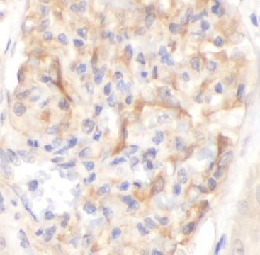
Immunohistochemistry of paraffin-embedded human kidney using FNab00129(ACVR1B antibody) at dilution of 1:100

various lysates were subjected to SDS PAGE followed by western blot with FNab00129(ACVR1B Antibody) at dilution of 1:1000
Share


