| TARGET | |
|---|---|
| SYNONYMS |
MIC-A; PERB11.1 |
| HOST SPECIES |
Rabbit |
| DESCRIPTION |
Anti-MICA antibody(DM157); Rabbit mAb |
| DELIVERY |
In Stock |
| UNIPROT ID |
Q29983 |
| IGG TYPE |
Rabbit IgG |
| CLONALITY |
Monoclonal |
| REACTIVITY |
Human |
| APPLICATIONS |
ELISA; Flow Cyt |
| RECOMMENDED DILUTIONS |
ELISA 1:5000-10000; Flow Cyt 1:100 |
| PURIFICATION |
Purified from cell culture supernatant by affinity chromatography |
| FORMULATION & RECONSTITUTION |
Lyophilized from sterile PBS, pH 7.4. Normally 5 % – 8% trehalose is added as protectants before lyophilization. Please see Certificate of Analysis for specific instructions of reconstitution. |
| STORAGE & SHIPPING |
Store at -20°C to -80°C for 12 months in lyophilized form. After reconstitution, if not intended for use within a month, aliquot and store at -80°C (Avoid repeated freezing and thawing). Lyophilized proteins are shipped at ambient temperature. |
| BACKGROUND |
This gene encodes the highly polymorphic major histocompatability complex class I chain-related protein A. The protein product is expressed on the cell surface; although unlike canonical class I molecules it does not seem to associate with beta-2-microglobulin. It is a ligand for the NKG2-D type II integral membrane protein receptor. The protein functions as a stress-induced antigen that is broadly recognized by intestinal epithelial gamma delta T cells. Variations in this gene have been associated with susceptibility to psoriasis 1 and psoriatic arthritis; and the shedding of MICA-related antibodies and ligands is involved in the progression from monoclonal gammopathy of undetermined significance to multiple myeloma. Alternative splicing of this gene results in multiple transcript variants. [provided by RefSeq; Jan 2014] |
| USAGE |
Research use only |
1
/
의
1
Dima Biotech
SKU(재고 관리 코드):DME100157
Anti-MICA antibody(DM157), Rabbit mAb
Anti-MICA antibody(DM157), Rabbit mAb
PRODUCT DATA
IMAGES

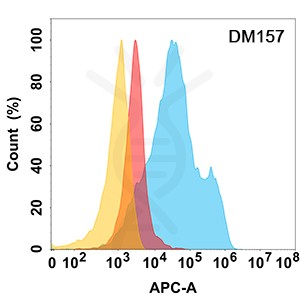
Figure 1. MICA protein is highly expressed on the surface of Expi293 cell membrane. Flow cytometry analysis with Anti-MICA (DM157) on Expi293 cells transfected with human MICA (Blue histogram) or Expi293 transfected with irrelevant protein (Red histogram), and Isotype antibody on Expi293 transfected with irrelevant protein (Orange histogram).
Share


