Western Blot: 1/500 - 1/2000. Immunohistochemistry: 1/100 - 1/300. Immunofluorescence: 1/200 - 1/1000. ELISA: 1/10000. Not yet tested in other applications.
Immunogen:
The antiserum was produced against synthesized peptide derived from human CA5B. AA range:241-290
Storage:
Rabbit
Storage:
-20°C/1 year
Clonality:
Polyclonal
Isotype:
IgG
Concentration:
1 mg/ml
Observed Band:
38kD
GeneID(Human):
11238
Human Swiss-Prot No:
Q9Y2D0
Cellular localization:
Mitochondrion.
Background:
Carbonic anhydrases (CAs) are a large family of zinc metalloenzymes that catalyze the reversible hydration of carbon dioxide. They participate in a variety of biological processes, including respiration, calcification, acid-base balance, bone resorption, and the formation of aqueous humor, cerebrospinal fluid, saliva, and gastric acid. They show extensive diversity in tissue distribution and in their subcellular localization. CA VB is localized in the mitochondria and shows the highest sequence similarity to the other mitochondrial CA, CA VA. It has a wider tissue distribution than CA VA, which is restricted to the liver. The differences in tissue distribution suggest that the two mitochondrial carbonic anhydrases evolved to assume different physiologic roles. [provided by RefSeq, Jul 2008],


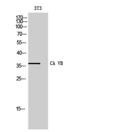 Western Blot analysis of 3T3 cells using CA VB Polyclonal Antibody
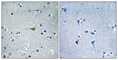 Immunohistochemical analysis of paraffin-embedded Human brain. Antibody was diluted at 1:100(4° overnight). High-pressure and temperature Tris-EDTA,pH8.0 was used for antigen retrieval. Negetive contrl (right) obtaned from antibody was pre-absorbed by i
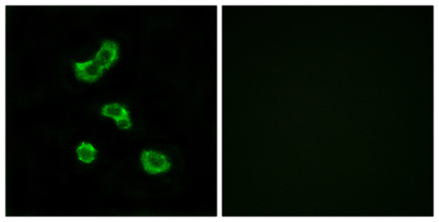 Immunofluorescence analysis of MCF7 cells, using CA5B Antibody. The picture on the right is blocked with the synthesized peptide.
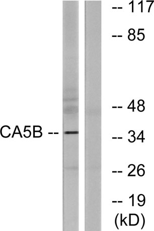 Western blot analysis of lysates from NIH/3T3 cells, using CA5B Antibody. The lane on the right is blocked with the synthesized peptide.