Tight junctions represent one mode of cell-to-cell adhesion in epithelial or endothelial cell sheets, forming continuous seals around cells and serving as a physical barrier to prevent solutes and water from passing freely through the paracellular space. These junctions are comprised of sets of continuous networking strands in the outwardly facing cytoplasmic leaflet, with complementary grooves in the inwardly facing extracytoplasmic leaflet. This gene encodes a component of tight junction strands, which is a member of the claudin family. The protein is an integral membrane protein and is one of the entry cofactors for hepatitis C virus. The gene methylation may be involved in esophageal tumorigenesis. This gene is adjacent to another family member CLDN9 on chromosome 16.[provided by RefSeq, Aug 2010],


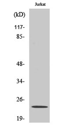 Western Blot analysis of various cells using Claudin-6 Polyclonal Antibody
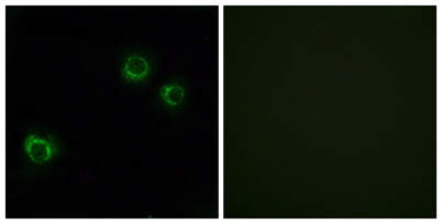 Immunofluorescence analysis of HUVEC cells, using CLDN6 Antibody. The picture on the right is blocked with the synthesized peptide.
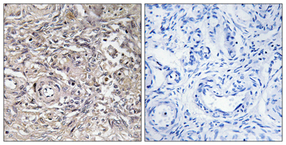 Immunohistochemistry analysis of paraffin-embedded human ovary tissue, using CLDN6 Antibody. The picture on the right is blocked with the synthesized peptide.
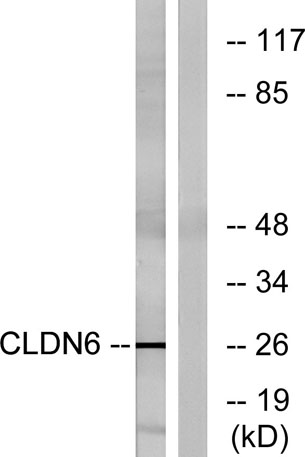 Western blot analysis of lysates from Jurkat cells, using CLDN6 Antibody. The lane on the right is blocked with the synthesized peptide.