MAP3K8; COT; ESTF; Mitogen-activated protein kinase kinase kinase 8; Cancer Osaka thyroid oncogene; Proto-oncogene c-Cot; Serine/threonine-protein kinase cot; Tumor progression locus 2; TPL-2
Applications:
WB;IHC;IF;ELISA
Recommended Dilutions:
Western Blot: 1/500 - 1/2000. Immunohistochemistry: 1/100 - 1/300. Immunofluorescence: 1/200 - 1/1000. ELISA: 1/40000. Not yet tested in other applications.
Immunogen:
The antiserum was produced against synthesized peptide derived from human COT. AA range:256-305
Storage:
Rabbit
Storage:
-20°C/1 year
Clonality:
Polyclonal
Isotype:
IgG
Concentration:
1 mg/ml
Observed Band:
60kD
GeneID(Human):
1326
Human Swiss-Prot No:
P41279
Cellular localization:
Cytoplasm .
Background:
This gene is an oncogene that encodes a member of the serine/threonine protein kinase family. The encoded protein localizes to the cytoplasm and can activate both the MAP kinase and JNK kinase pathways. This protein was shown to activate IkappaB kinases, and thus induce the nuclear production of NF-kappaB. This protein was also found to promote the production of TNF-alpha and IL-2 during T lymphocyte activation. This gene may also utilize a downstream in-frame translation start codon, and thus produce an isoform containing a shorter N-terminus. The shorter isoform has been shown to display weaker transforming activity. Alternate splicing results in multiple transcript variants that encode the same protein. [provided by RefSeq, Sep 2011],


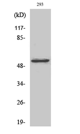 Western Blot analysis of various cells using COT Polyclonal Antibody
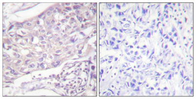 Immunohistochemical analysis of paraffin-embedded Human breast cancer. Antibody was diluted at 1:100(4° overnight). High-pressure and temperature Tris-EDTA,pH8.0 was used for antigen retrieval. Negetive contrl (right) obtaned from antibody was pre-absor
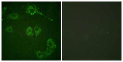 Immunofluorescence analysis of HUVEC cells, using COT Antibody. The picture on the right is blocked with the synthesized peptide.
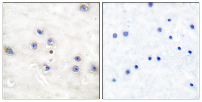 Immunohistochemistry analysis of paraffin-embedded human brain tissue, using COT Antibody. The picture on the right is blocked with the synthesized peptide.