

Cusabio
SKU(재고 관리 코드):CSB-MA281265
PHGDH Monoclonal Antibody
PHGDH Monoclonal Antibody
가격·납기 및 기술자료를 확인해 드립니다.
제품 규격과 수량을 선택한 후 견적을 요청해 주세요. 대체품 검토와 SDS·COA·데이터시트 요청도 함께 접수할 수 있습니다.
이 제품 견적 요청대표번호 070-8028-3500 · info@rndmate.com
| Release Date | 2025-12-04 |
|---|---|
| Product Code | CSB-MA281265 |
| Short Name | PHGDH Monoclonal Antibody |
| Antibody Type | Monoclonal Antibody |
| List Price | $350/100ul, $210/50ul |
| Species Reactivity | Human, Mouse, Rat |
| Tested Applications | ELISA, WB, FC, ICC, IHC |
| Dilution | WB:1:500-1:2500, FC:1:200-1:2000, ICC:1:100-1:1000, IHC:1:100-1:200 |
| Purification Method | Affinity chromatography |
| Buffer Formulation | PBS, 0.02% Sodium Azide, 50% Glycerol, pH 7.4. |
| Antigen | Recombinant Human PHGDH protein |
| Antigen Species | Human |
| Uniprot ID | O43175 |
| Target Name | PHGDH |
| Clonality | Monoclonal |
| Clone ID | 4G2 |
| Host Species | Mouse |
| Isotype | IgG2a |
| Conjugation | Non-conjugated |
| Form | Liquid |
| Storage Condition | Upon delivery aliquot and store at -20°C or -80°C. Avoid repeated freeze. |
| TDS URL | https://www.cusabio.com/datasheet/12947827/ |
| Size | 50ul |
Image 1
Flow cytometric analysis of Phosphoglycerate dehydrogenase expression in C2C12 cells using Phosphoglycerate dehydrogenase antibody. Green, isotype control; red, Phosphoglycerate dehydrogenase.
Image 2
Immunocytochemical staining of C2C12 cells with Phosphoglycerate dehydrogenase antibody. Nuclei were stained blue with DAPI; Phosphoglycerate dehydrogenase was stained magenta with Alexa Fluor® 647. Images were taken using Leica stellaris 5. Protein abundance based on laser Intensity and smart gain: Medium. Scale bar, 20 μm.
Image 3
Immunohistochemistry was performed on paraffin-embedded human glioblastoma using phosphoglycerate dehydrogenase antibody. Antigen retrieval was done in sodium citrate buffer (pH 6.0). DAB was used for detection, with hematoxylin counterstaining. Images were acquired using a Nikon Ci-L Plus microscope (40× objective). Scale bar: 25 μm.
Image 4
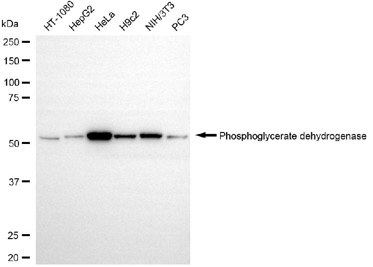
Western blotting analysis using phosphoglycerate dehydrogenase antibody. Total cell lysates (30 μg) from various cell lines were loaded and separated by SDS-PAGE. The blot was incubated with phosphoglycerate dehydrogenase antibody and HRP-conjugated goat anti-mouse secondary antibody respectively.
Share