Histones are basic nuclear proteins that are responsible for the nucleosome structure of the chromosomal fiber in eukaryotes. Two molecules of each of the four core histones (H2A, H2B, H3, and H4) form an octamer, around which approximately 146 bp of DNA is wrapped in repeating units, called nucleosomes. The linker histone, H1, interacts with linker DNA between nucleosomes and functions in the compaction of chromatin into higher order structures. This gene is intronless and encodes a replication-dependent histone that is a member of the histone H4 family. Transcripts from this gene lack polyA tails but instead contain a palindromic termination element. This gene is found in the histone microcluster on chromosome 6p21.33. [provided by RefSeq, Aug 2015],


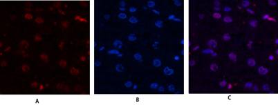 Immunofluorescence analysis of human-liver tissue. 1,Histone H4 Polyclonal Antibody(red) was diluted at 1:200(4° overnight). 2, Cy3 labled Secondary antibody was diluted at 1:300(room temperature, 50min).3, Picture B: DAPI(blue) 10min. Picture A:Target. P
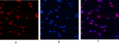 Immunofluorescence analysis of rat-heart tissue. 1,Histone H4 Polyclonal Antibody(red) was diluted at 1:200(4° overnight). 2, Cy3 labled Secondary antibody was diluted at 1:300(room temperature, 50min).3, Picture B: DAPI(blue) 10min. Picture A:Target. Pic
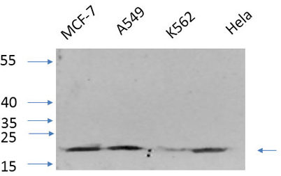 Western Blot analysis of various cells using primary antibody diluted at 1:1000(4°C overnight). Secondary antibody:Goat Anti-rabbit IgG IRDye 800( diluted at 1:5000, 25°C, 1 hour). cells nucleus extracted by Minute TM Cytoplasmic and Nuclear Fractionatio
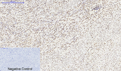 Immunohistochemical analysis of paraffin-embedded Human-uterus tissue. 1,Histone H4 Polyclonal Antibody was diluted at 1:200(4°C,overnight). 2, Sodium citrate pH 6.0 was used for antibody retrieval(>98°C,20min). 3,Secondary antibody was diluted at 1:200(r