| Product name: |
LATS1/2 antibody |
|
| Alternative Names: |
Serine/threonine-protein kinase LATS1 (EC 2.7.11.1) (Large tumor suppressor homolog 1) (WARTS protein kinase) (h-warts) |
| Applications: |
WB;ELISA;IHC |
| Recommended Dilutions: |
WB 1:500-2000;IHC-p 1:50-300; ELISA 2000-20000 |
| Immunogen: |
Synthesized peptide derived from human LATS1/2 AA range: 1050-1130 |
| Storage: |
Rabbit |
| Storage: |
-20°C/1 year |
| Clonality: |
Polyclonal |
| Isotype: |
IgG |
| Concentration: |
1 mg/ml |
| Observed Band: |
140kD |
| GeneID(Human): |
9113 |
| Human Swiss-Prot No: |
O95835 |
| Cellular localization: |
Cytoplasm, cytoskeleton, microtubule organizing center, centrosome . Cytoplasm, cytoskeleton, spindle . Midbody . Cytoplasm, cytoskeleton, microtubule organizing center, spindle pole body . Localizes to the centrosomes throughout interphase but migrates to the mitotic apparatus, including spindle pole bodies, mitotic spindle, and midbody, during mitosis. . |
| Background: |
The protein encoded by this gene is a putative serine/threonine kinase that localizes to the mitotic apparatus and complexes with cell cycle controller CDC2 kinase in early mitosis. The protein is phosphorylated in a cell-cycle dependent manner, with late prophase phosphorylation remaining through metaphase. The N-terminal region of the protein binds CDC2 to form a complex showing reduced H1 histone kinase activity, indicating a role as a negative regulator of CDC2/cyclin A. In addition, the C-terminal kinase domain binds to its own N-terminal region, suggesting potential negative regulation through interference with complex formation via intramolecular binding. Biochemical and genetic data suggest a role as a tumor suppressor. This is supported by studies in knockout mice showing development of soft-tissue sarcomas, ovarian stromal cell tumors and a high sensitivity to carcinogenic treatmen |
| Species Reactivity: |
Human;Mouse;Rat |


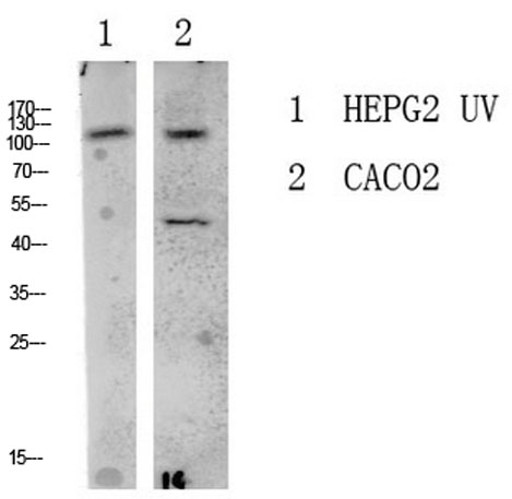 Western blot analysis of various lysate, antibody was diluted at 1000. Secondary antibody(catalog#:RS0002) was diluted at 1:20000
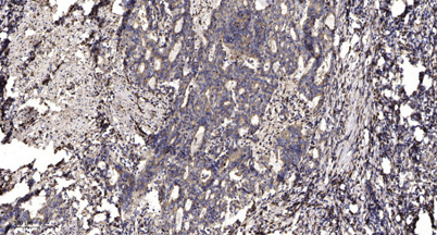 Immunohistochemical analysis of paraffin-embedded human Gastric adenocarcinoma. 1, Antibody was diluted at 1:200(4° overnight). 2, Tris-EDTA,pH9.0 was used for antigen retrieval. 3,Secondary antibody was diluted at 1:200(room temperature, 45min).