DUSP6; MKP3; PYST1; Dual specificity protein phosphatase 6; Dual specificity protein phosphatase PYST1; Mitogen-activated protein kinase phosphatase 3; MAP kinase phosphatase 3; MKP-3
Applications:
WB;IHC;IF;ELISA
Recommended Dilutions:
Western Blot: 1/500 - 1/2000. Immunohistochemistry: 1/100 - 1/300. Immunofluorescence: 1/200 - 1/1000. ELISA: 1/20000. Not yet tested in other applications.
Immunogen:
The antiserum was produced against synthesized peptide derived from human DUSP6. AA range:61-110
Storage:
Rabbit
Storage:
-20°C/1 year
Clonality:
Polyclonal
Isotype:
IgG
Concentration:
1 mg/ml
Observed Band:
42kD
GeneID(Human):
1848
Human Swiss-Prot No:
Q16828
Cellular localization:
Cytoplasm .
Background:
The protein encoded by this gene is a member of the dual specificity protein phosphatase subfamily. These phosphatases inactivate their target kinases by dephosphorylating both the phosphoserine/threonine and phosphotyrosine residues. They negatively regulate members of the mitogen-activated protein (MAP) kinase superfamily (MAPK/ERK, SAPK/JNK, p38), which are associated with cellular proliferation and differentiation. Different members of the family of dual specificity phosphatases show distinct substrate specificities for various MAP kinases, different tissue distribution and subcellular localization, and different modes of inducibility of their expression by extracellular stimuli. This gene product inactivates ERK2, is expressed in a variety of tissues with the highest levels in heart and pancreas, and unlike most other members of this family, is localized in the cytoplasm. Mutations in t


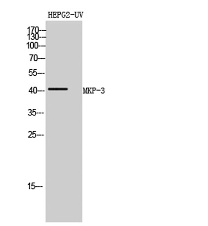 Western Blot analysis of HEPG2-UV cells using MKP-3 Polyclonal Antibody diluted at 1:500
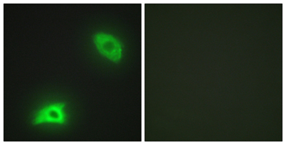 Immunofluorescence analysis of HeLa cells, using DUSP6 Antibody. The picture on the right is blocked with the synthesized peptide.
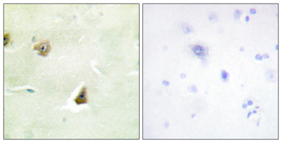 Immunohistochemistry analysis of paraffin-embedded human brain tissue, using DUSP6 Antibody. The picture on the right is blocked with the synthesized peptide.