Western Blot: 1/500 - 1/2000. Immunohistochemistry: 1/100 - 1/300. ELISA: 1/10000. Not yet tested in other applications.
Immunogen:
The antiserum was produced against synthesized peptide derived from human PSMD11. AA range:271-320
Storage:
Rabbit
Storage:
-20°C/1 year
Clonality:
Polyclonal
Isotype:
IgG
Concentration:
1 mg/ml
Observed Band:
42kD
GeneID(Human):
5717
Human Swiss-Prot No:
O00231
Cellular localization:
Nucleus . Cytoplasm, cytosol .
Background:
The 26S proteasome is a multicatalytic proteinase complex with a highly ordered structure composed of 2 complexes, a 20S core and a 19S regulator. The 20S core is composed of 4 rings of 28 non-identical subunits; 2 rings are composed of 7 alpha subunits and 2 rings are composed of 7 beta subunits. The 19S regulator is composed of a base, which contains 6 ATPase subunits and 2 non-ATPase subunits, and a lid, which contains up to 10 non-ATPase subunits. Proteasomes are distributed throughout eukaryotic cells at a high concentration and cleave peptides in an ATP/ubiquitin-dependent process in a non-lysosomal pathway. This gene encodes a member of the proteasome subunit S9 family that functions as a non-ATPase subunit of the 19S regulator and is phosphorylated by AMP-activated protein kinase. Alternatively spliced transcript variants have been observed for this gene. [provided by RefSeq,


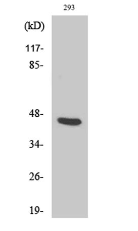 Western Blot analysis of various cells using PSMD11 Polyclonal Antibody
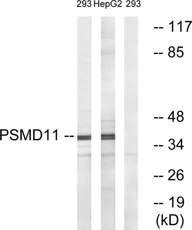 Western blot analysis of lysates from 293 and HepG2 cells, using PSMD11 Antibody. The lane on the right is blocked with the synthesized peptide.
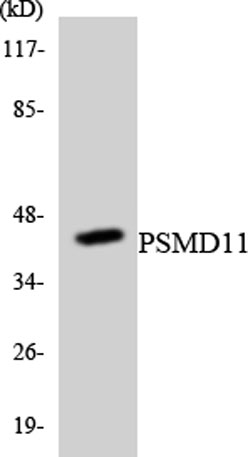 Western blot analysis of the lysates from RAW264.7cells using PSMD11 antibody.
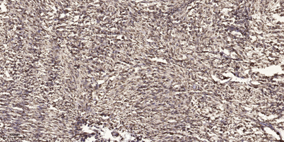 Immunohistochemical analysis of paraffin-embedded human small intestinal carcinoma tissue. 1,primary Antibody was diluted at 1:200(4° overnight). 2, Sodium citrate pH 6.0 was used for antigen retrieval(>98°C,20min). 3,Secondary antibody was diluted at 1:2