Western Blot: 1/500 - 1/2000. Immunohistochemistry: 1/100 - 1/300. Immunofluorescence: 1/200 - 1/1000. ELISA: 1/20000. Not yet tested in other applications.
Immunogen:
The antiserum was produced against synthesized peptide derived from human PTTG1. AA range:111-160
Storage:
-20°C/1 year
Storage:
Rabbit
Clonality:
Polyclonal
Isotype:
IgG
Concentration:
1 mg/ml
Observed Band:
30kD
GeneID(Human):
9232
Human Swiss-Prot No:
O95997
Cellular localization:
Cytoplasm. Nucleus.
Background:
The encoded protein is a homolog of yeast securin proteins, which prevent separins from promoting sister chromatid separation. It is an anaphase-promoting complex (APC) substrate that associates with a separin until activation of the APC. The gene product has transforming activity in vitro and tumorigenic activity in vivo, and the gene is highly expressed in various tumors. The gene product contains 2 PXXP motifs, which are required for its transforming and tumorigenic activities, as well as for its stimulation of basic fibroblast growth factor expression. It also contains a destruction box (D box) that is required for its degradation by the APC. The acidic C-terminal region of the encoded protein can act as a transactivation domain. The gene product is mainly a cytosolic protein, although it partially localizes in the nucleus. Three transcript variants encoding the same protein have been fo


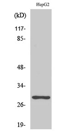 Western Blot analysis of various cells using PTTG1/2/3 Polyclonal Antibody diluted at 1:1000
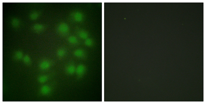 Immunofluorescence analysis of HUVEC cells, using PTTG1 Antibody. The picture on the right is blocked with the synthesized peptide.
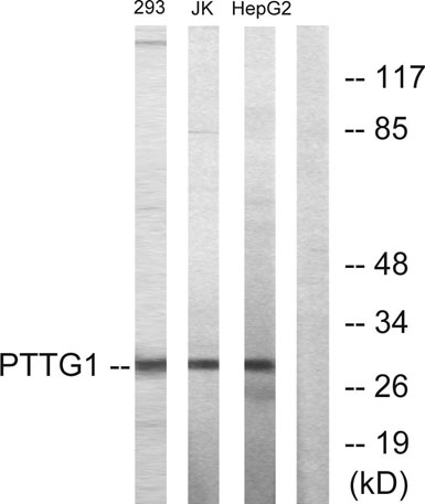 Western blot analysis of lysates from HepG2, Jurkat, and 293 cells, using PTTG1 Antibody. The lane on the right is blocked with the synthesized peptide.
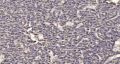 Immunohistochemical analysis of paraffin-embedded human liver cancer. 1, Antibody was diluted at 1:200(4° overnight). 2, Tris-EDTA,pH9.0 was used for antigen retrieval. 3,Secondary antibody was diluted at 1:200(room temperature, 45min).